

close
Choose Your Site
Global
Social Media
Views: 0 Author: Site Editor Publish Time: 2026-08-26 Origin: Site


Formulating with salicylic acid presents well-documented challenges regarding aqueous solubility and skin irritation. While cyclodextrin encapsulation offers a theoretical solution, verifying that a true inclusion complex has formed—rather than a mere physical mixture—is a technical hurdle you cannot bypass. Utilizing unverified complexes leads to formulation instability, crystallization over time, and failure to mitigate the irritation profile of the active ingredient. Procurement and R&D teams must rely on definitive analytical data to validate raw materials before scaling up production. Establishing a rigorous analytical testing framework is essential for quality control. This guide details the specific spectroscopic, thermal, morphological, and solubility tests required to definitively confirm the formation of a true inclusion complex within the Salicylic Acid Series, ensuring product efficacy, long-term shelf stability, and consumer safety.
Solid-State vs. Solution-State Verification: Complete confirmation requires a combination of solid-state analysis (FT-IR, DSC, XRD, SEM) and solution-state analysis (NMR, Fluorescence Spectroscopy).
The Gold Standards: Nuclear Magnetic Resonance (1H NMR and 13C NMR) and Differential Scanning Calorimetry (DSC) provide the most definitive evidence of guest-host molecular interactions.
Quality Control vs. R&D: While NMR is critical during the initial R&D and structural elucidation phase, FT-IR and UV-Vis spectroscopy serve as more practical, scalable methods for routine batch-to-batch quality control.
Formulation Impact: Verified inclusion complexes directly correlate to the successful development of stable, high concentration salicylic acid products with enhanced water solubility, improved skin permeability, and controlled release profiles.
Standard salicylic acid suffers from severe physical limitations in aqueous environments. It has a pKa of 2.97, meaning at typical skin pH levels (4.5 to 5.5), it remains largely unionized. While this unionized state favors skin penetration, it results in an abysmal water solubility limit of approximately 0.2% at room temperature. To formulate a standard 2% exfoliating serum, chemists typically rely on high concentrations of ethanol, propylene glycol, or other harsh solvents. These solvents strip the epidermal barrier, exacerbating the inherent irritation potential of the acid. Formulators require a functional complex that delivers enhanced solubility, protects the active sites from premature degradation, and provides a sustained release mechanism upon application.
The solution lies in the precise mechanism of the cyclodextrin salicylic acid complex. Beta-cyclodextrin features a unique truncated cone structure with an internal cavity diameter of roughly 6.0 to 6.5 Å. This hydrophobic interior cavity perfectly accommodates the lipophilic benzene ring of the salicylic acid molecule. Simultaneously, the hydrophilic exterior of the cyclodextrin shell ensures excellent compatibility with the surrounding water phase. This molecular encapsulation shields the acidic functional groups from direct contact with the skin surface upon initial application, drastically reducing erythema while maintaining keratolytic efficacy.
Manufacturing variables introduce significant risk during production. Preparation techniques heavily impact complexation efficiency. Common methods include the kneading method, co-precipitation, solvent evaporation, and spray drying. Temperature fluctuations, mixing RPMs, and solvent-to-water ratios alter the final yield of the complex. Because these manufacturing methods introduce inherent variability, rigorous analytical testing is mandatory. You cannot assume encapsulation has occurred simply because the raw materials were mixed in a tank.
Utilizing unverified materials carries massive implementation risks. A simple physical mixture of salicylic acid and cyclodextrin will fail under formulation stress. When added to an aqueous serum or toner, the unencapsulated salicylic acid will rapidly precipitate out of solution. This leads to several catastrophic manufacturing failures:
Stratification of the active ingredient in the holding hopper before filling.
Visible crystallization and sediment formation at the bottom of the final packaging.
Clogging of inline micron filters during the bottling process.
Severe consumer safety risks due to uneven active distribution, causing chemical burns from concentrated acid pockets.
Validating the complex ensures you are purchasing a functional raw material rather than a simple, unstable blend.
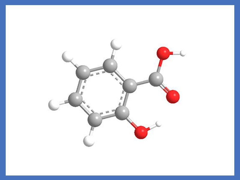
The objective of primary analytical testing is to definitively prove molecular encapsulation. We must transition from merely observing bulk physical properties to precisely measuring chemical interactions at the molecular level. The following techniques provide the foundational proof of complexation.
Nuclear Magnetic Resonance spectroscopy evaluates the exact insertion mechanism of the guest molecule. 1H NMR and 13C NMR detect the spatial arrangement of the salicylic acid molecule within the cyclodextrin cavity. When the guest enters the host cavity, the magnetic environment of the atoms changes, leading to measurable shifts in the NMR spectrum. For this analysis, samples are typically dissolved in D2O or DMSO-d6 and analyzed using a 400 MHz or 600 MHz spectrometer.
Data interpretation focuses on observing chemical shift changes (Δδ). Analysts specifically look for shielding or deshielding effects on the inner cavity protons of the beta-cyclodextrin. The H-3 and H-5 protons are located inside the cyclodextrin cavity. If salicylic acid has successfully entered the cavity, the anisotropic shielding from the aromatic ring of the salicylic acid causes these specific protons to exhibit significant upfield shifts (lower ppm values) compared to an empty cyclodextrin molecule. Protons on the exterior of the cyclodextrin (H-1, H-2, H-4) typically show little to no shift.
| Proton Location (Beta-Cyclodextrin) | Position Relative to Cavity | Expected NMR Shift (Δδ) upon Complexation |
|---|---|---|
| H-3 | Inside (Wide edge) | Significant upfield shift |
| H-5 | Inside (Narrow edge) | Significant upfield shift |
| H-1, H-2, H-4 | Exterior surface | Negligible shift (< 0.01 ppm) |
Furthermore, 2D ROESY (Rotating-frame Overhauser Effect Spectroscopy) NMR detects spatial proximity between the aromatic protons of the salicylic acid and the inner cavity protons of the cyclodextrin. Cross-peaks in a ROESY spectrum indicate that two protons are within 4 Å of each other, providing absolute proof of insertion. NMR stands as the undisputed gold standard for proving inclusion in the solution state. It determines the exact geometry and stoichiometry of the complex in aqueous environments.
FT-IR spectroscopy monitors changes in the vibrational frequencies of specific functional groups. When molecules interact, their chemical bonds absorb infrared light at different frequencies. This technique is highly sensitive to hydrogen bonding and changes in the solid-state environment. Samples are typically prepared using the KBr pellet method, where 1-2 mg of the complex is ground with 100 mg of anhydrous potassium bromide and pressed under 10 tons of force to form a transparent disk.
Data interpretation requires focusing on the carbonyl (C=O) and hydroxyl (-OH) stretching bands of salicylic acid. Pure salicylic acid displays a sharp, distinct carbonyl stretch around 1655 cm⁻¹. In a true salicylic acid inclusion complex, these bands will shift, broaden, or disappear entirely. This phenomenon occurs due to the restriction of the guest molecule within the cyclodextrin cavity and the breaking of the original crystalline hydrogen bonds. The cyclodextrin's massive hydroxyl bands (typically spanning 3200 to 3400 cm⁻¹) often mask the guest's signals, further indicating successful solid-state dispersion.
FT-IR serves as a highly reliable, cost-effective method for solid-state confirmation. It easily differentiates a true inclusion complex from a physical mixture. A physical mixture will simply display a superimposition of the individual spectra of both raw materials without any significant band shifts or peak broadening.
DSC outlines the thermal analysis techniques used to evaluate the melting, boiling, and sublimation points of a compound. It measures the heat flow into or out of a sample as it is heated, cooled, or held at a constant temperature, revealing phase transitions. For this test, 3 to 5 mg of the sample is placed in a sealed aluminum pan and heated from 25°C to 300°C at a rate of 10°C per minute under a constant nitrogen purge (50 mL/min).
Pure crystalline salicylic acid exhibits a distinct, sharp endothermic melting peak typically around 159°C. In a successfully formed complex, this characteristic melting peak disappears completely or shifts significantly. The disappearance of the melting peak indicates that the salicylic acid is molecularly dispersed within the cyclodextrin cavity and no longer exists in its crystalline state. A physical mixture will still display the endothermic peak of the free active, albeit potentially reduced in size proportional to its concentration in the blend.
To properly interpret DSC data, follow these analytical steps:
Run a baseline scan of pure, empty beta-cyclodextrin to identify its broad dehydration endotherm (typically around 80-100°C).
Run a scan of pure salicylic acid to confirm the sharp melting endotherm at 159°C.
Run a scan of a simple 1:1 physical mixture. You must observe the 159°C peak to validate the method's sensitivity.
Run the manufactured inclusion complex. The complete absence of the 159°C peak confirms 100% amorphization and successful encapsulation.
While NMR and DSC provide primary proof, supplementary tests support this data, quantify the complexation efficiency, and evaluate the physical characteristics of the resulting material. These secondary methods are essential for complete material characterization and formulation scaling.
The Higuchi and Connors method is the standard protocol for evaluating phase solubility. This test involves adding an excess amount of salicylic acid to 20 mL glass vials containing aqueous solutions with increasing concentrations of cyclodextrin (e.g., 0 to 10 mM). The suspensions are sealed and shaken at 100 RPM in a temperature-controlled water bath at 25°C for 48 hours until equilibrium is reached. The samples are then filtered through a 0.45-micron syringe filter and analyzed for active concentration using HPLC.
Plotting the concentration of dissolved water soluble salicylic acid against the increasing concentrations of cyclodextrin yields a phase solubility diagram. Typically, this results in an AL-type curve, where the solubility of the guest increases linearly with the host concentration. This linear relationship indicates a 1:1 stoichiometric ratio.
This test is essential for calculating the stability constant (Kc) of the complex using the slope of the phase solubility diagram. The stability constant dictates the complex's behavior in aqueous formulations. A value between 100 and 1000 M⁻¹ is generally considered ideal. A value too low means the complex will easily dissociate in the bottle, while a value too high means the active will not release onto the skin upon application.
XRD details the assessment of the crystallinity of the solid material. It measures the diffraction of X-rays by the crystal lattice of the sample. The instrument typically uses Cu K-alpha radiation operated at 40 kV and 30 mA, scanning the sample from 5 to 40 degrees 2-theta with a step size of 0.02 degrees.
Data interpretation involves contrasting the diffractograms of the raw materials with the final product. Pure, crystalline salicylic acid displays numerous sharp, intense diffraction peaks at specific 2-theta angles (e.g., 11.0°, 15.5°, 17.2°). In contrast, an amorphous cyclodextrin inclusion complex exhibits a broad, diffuse halo with the complete absence of the guest's characteristic sharp peaks.
XRD is highly useful for confirming the complete amorphization of the guest molecule. It perfectly supports DSC findings, confirming that the crystalline lattice of the active has been completely disrupted through encapsulation. If sharp peaks remain in the complex's diffractogram, it indicates unencapsulated, free salicylic acid is still present in the batch.
SEM utilizes high-resolution microscopic examinations to visually assess the surface morphology and particle size of the material. Samples are mounted on double-sided carbon tape and sputter-coated with a thin layer of gold or palladium for 60 seconds to enhance conductivity and prevent charge buildup before imaging under a vacuum at an accelerating voltage of 10 to 15 kV.
The visual contrast is striking. Pure salicylic acid presents as distinct, needle-like crystalline structures. Pure cyclodextrin often appears as blocky, irregular, or spherical crystals depending on its manufacturing origin. A successfully formed cyclodextrin salicylic acid complex reveals amorphous, irregular aggregates that look entirely different from either starting material. The original needle-like structures completely vanish, replaced by a homogenous, fluffy powder morphology.
SEM provides compelling visual confirmation of solid-state changes. It acts as a powerful complement to DSC and XRD data, offering a tangible look at the physical transformation resulting from the inclusion process.
The microenvironment inside the cyclodextrin cavity significantly affects the absorption and emission of light by the guest molecule. The cavity is relatively non-polar compared to the bulk aqueous environment.
Transferring salicylic acid from a highly polar aqueous environment into the non-polar cyclodextrin cavity results in measurable changes. Analysts observe shifts in the maximum absorption wavelength (lambda max) or changes in molar absorptivity. Because the cavity restricts the rotational freedom of the guest and shields it from quenching agents in the solvent, fluorescence intensity often increases significantly upon complexation. This provides rapid, measurable proof of inclusion in the solution state and can be used as a quick assay method during production.
Bridging the gap between analytical chemistry and practical formulation performance requires detailing tests that confirm biological and physical efficacy. Analytical data proves the structure exists; performance testing proves the structure functions as intended on human skin.
In vitro release and permeability studies utilize Franz diffusion cell methods. These setups measure the controlled release rate and enhanced skin permeability of the complex compared to free salicylic acid. A synthetic membrane (like Strat-M) or human cadaver skin separates the donor compartment (containing the complex formulation) from the receptor compartment. The receptor compartment is filled with a buffer, such as PBS pH 7.4 with 4% BSA, to maintain sink conditions. The receptor fluid is sampled at specific intervals (e.g., 1, 2, 4, 8, 12, and 24 hours).
A verified complex will show a sustained, steady release profile, avoiding the rapid spike in concentration typical of unencapsulated acids. Furthermore, the enhanced water solubility allows for a higher concentration gradient in the donor compartment, directly improving epidermal permeability without requiring barrier-damaging solvents.
Skin surface concentration and irritation assays are equally critical. Clinical patch testing proves that the complex maintains an effective salicylic acid concentration at the skin surface. These assays measure trans-epidermal water loss (TEWL) using a Tewameter and erythema using a Mexameter. A true inclusion complex significantly reduces epidermal barrier disruption and redness compared to a standard physical mixture, proving its value in sensitive skin formulations.
Translating R&D success to routine quality control involves conceptual trade-offs. Rigorous R&D testing, including NMR, XRD, and SEM, is necessary for initial structural elucidation and supplier qualification. However, these methods are too slow, expensive, and complex for routine batch release on a manufacturing floor. A scalable QC framework must rely on faster, robust methods. FT-IR and UV-Vis spectroscopy serve as the ideal tools for daily batch validation. By comparing the FT-IR spectrum of every new batch against a validated reference standard, manufacturers can quickly confirm that the inclusion complex structure remains consistent.
| Analytical Method | Primary Application | Data Output | Scalability for QC |
|---|---|---|---|
| NMR Spectroscopy | R&D / Structural Proof | Proton shifts, spatial geometry | Low (High cost, complex prep) |
| DSC | R&D / Thermal Validation | Melting point disappearance | Medium (Requires specific equipment) |
| FT-IR Spectroscopy | Routine QC / Batch Release | Functional group band shifts | High (Fast, cost-effective) |
| UV-Vis Spectroscopy | Routine QC / Assay | Molar absorptivity changes | High (Standard lab equipment) |
Implementation risks center heavily on partial inclusion. In poorly controlled manufacturing processes, only a percentage of the active might be encapsulated, leaving a dangerous amount of free acid in the bulk powder. Mitigate this risk by establishing strict acceptable ranges for thermal shifts in DSC data and setting precise solubility thresholds in the Certificate of Analysis (CoA). If a batch fails to meet the minimum aqueous solubility threshold, it indicates a high presence of unencapsulated material.
Compliance and documentation form the final layer of defense. Request comprehensive analytical dossiers from suppliers of any high concentration salicylic acid product. A standard specification sheet is insufficient. You need access to the original spectra and thermal curves to ensure regulatory compliance and guarantee formulation safety across your entire product line.
Request a comprehensive Certificate of Analysis (CoA) containing DSC and FT-IR overlay graphs before approving any new raw material supplier for inclusion complexes.
Implement routine UV-Vis spectroscopy and phase solubility checks for every incoming batch to verify active concentration and complex stability.
Conduct Franz diffusion cell testing on pilot formulations to establish baseline release kinetics compared to unencapsulated salicylic acid controls.
Establish strict acceptable ranges for thermal shifts in your internal quality control documentation to prevent partial inclusion failures from reaching the production floor.
A: A physical mixture retains the individual chemical, morphological, and thermal properties of both components. The active remains crystalline and poorly soluble. An inclusion complex alters the guest molecule's physical state, completely disrupting its crystal lattice. This changes its melting point, spectroscopic signature, and drastically increases its aqueous solubility.
A: It eliminates the need for harsh solvents like high-concentration alcohol or glycols. This significantly reduces skin barrier disruption and erythema. Furthermore, the encapsulation enhances permeability and allows formulators to create stable, high-concentration aqueous serums without the risk of active crystallization over time.
A: While FT-IR is excellent for routine quality control and shows clear band shifts due to hydrogen bond disruption, it should not be the sole proof during initial R&D. Complete confirmation requires pairing FT-IR with thermal analysis (DSC) or solution-state analysis (NMR) to definitively prove molecular encapsulation.
A: The kneading method involves mixing the active and cyclodextrin with a small amount of solvent to form a paste. The mechanical shear forces and localized heating drive the guest into the cavity. Variations in kneading time, temperature, and solvent ratio directly impact the final yield and stability of the complex.
A: The sharp endothermic melting peak represents the energy required to break the crystalline lattice of pure salicylic acid. When successfully encapsulated, the acid molecules are isolated within the cyclodextrin cavities. They no longer form a crystal lattice, rendering the material amorphous and eliminating the characteristic melting peak.
A: Phase solubility determines the stability constant (Kc) of the complex. This value dictates how tightly the cyclodextrin holds the salicylic acid. An optimal Kc ensures the complex remains stable in the aqueous formulation but releases the active efficiently upon contact with the skin's surface.


